The cell surface is regulated by both membrane proteins and structural lipids that together control signaling, transport, adhesion and receptor organization. While surface proteomics identifies membrane-associated proteins, membrane lipidomics characterizes the lipid environment that shapes membrane structure and receptor localization.
Panome Bio’s Cell Surface Multi-Omics platform combines:
- Cell Surface Proteomics
- Cell Membrane Lipidomics
- Integrated protein–lipid analysis
From matched membrane fractions to study coordinated remodeling at the membrane interface.
KRAS Knockdown Remodels the Surface Proteome
Membrane-enriched fractions from HCT-116 KRAS knockdown and wild-type colorectal cancer cells were analyzed using Cell Surface Proteomics and untargeted Membrane Lipidomics.
Across all samples:
- 6,272 protein groups were detected
- 472 high-confidence surface proteins were retained following SPAT filtering
These proteins included:
- Surface receptors
- Transporters
- GPI-anchored proteins
- Adhesion-associated proteins
Surface Changes Concentrate in Transport and Signaling Proteins
Differential analysis identified 17 significantly altered cell surface proteins.
Reduced proteins included:
- FOLR1
- SLC7A11/xCT
- MCAM
- AREG
These proteins are associated with nutrient transport, redox regulation, and adhesion signaling.
Elevated proteins included:
- TACSTD2 (TROP2)
- FLOT1
- EGFL7
- SUSD2
Several of these proteins are associated with lipid raft organization, immune modulation, and therapeutic targeting in oncology.
Together, these shifts suggest coordinated restructuring of surface signaling and transport networks following KRAS suppression.
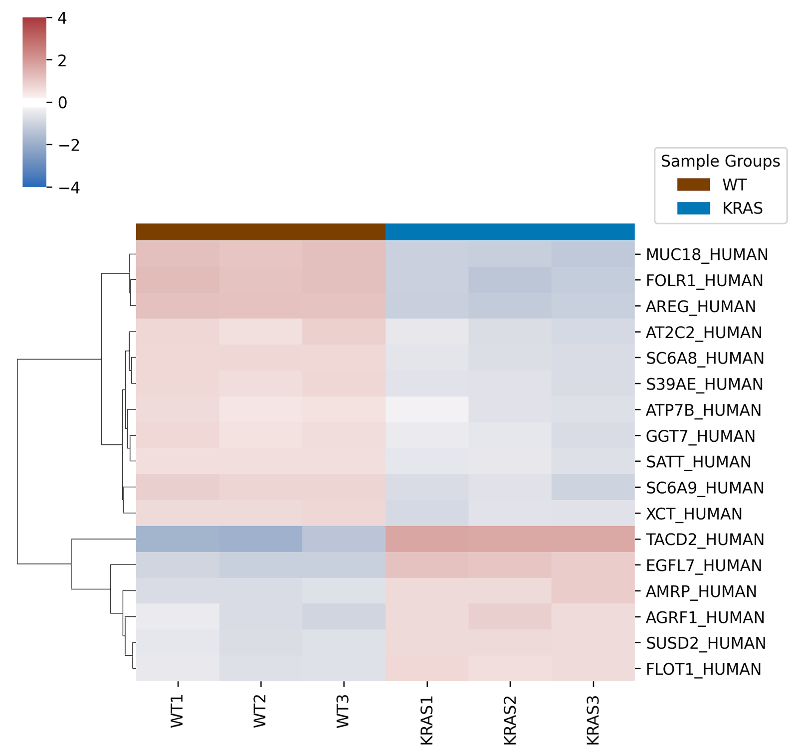
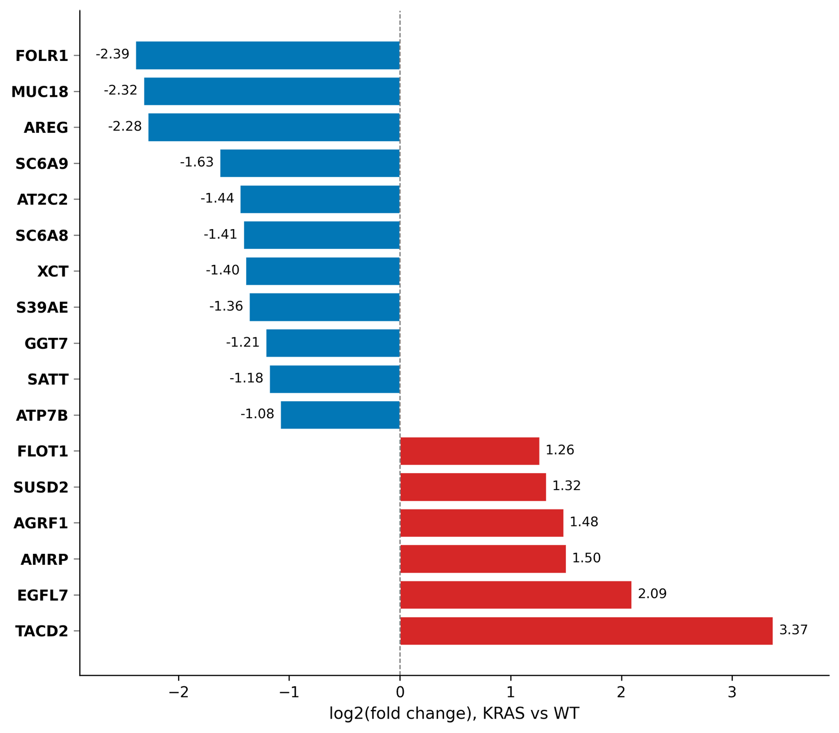
Surface Transport and Signaling Proteins Shift Following KRAS Knockdown
KRAS knockdown alters a defined subset of cell surface proteins associated with nutrient transport, adhesion, and signaling.
Heatmap clustering (right) shows clear separation between KRAS and wild-type samples, while fold-change analysis highlights coordinated increases and decreases across the surface proteome in the boxplot (right).
Membrane Lipidomics Reveals Structural Remodeling
Unlike conventional whole-cell lipidomics, membrane lipidomics enriches for lipids present at the cellular membrane interface, enabling direct investigation of membrane microdomains involved in receptor organization and signaling.
Untargeted membrane lipidomics identified:
- 5,749 lipid features
- 1,282 high-confidence lipids
- 73 significantly altered membrane lipids
KRAS knockdown cells showed broad remodeling across:
- Glycerophospholipids
- Sphingolipids
- Sterol-associated lipid classes
These lipid classes play central roles in membrane order, lipid raft organization, and receptor partitioning.
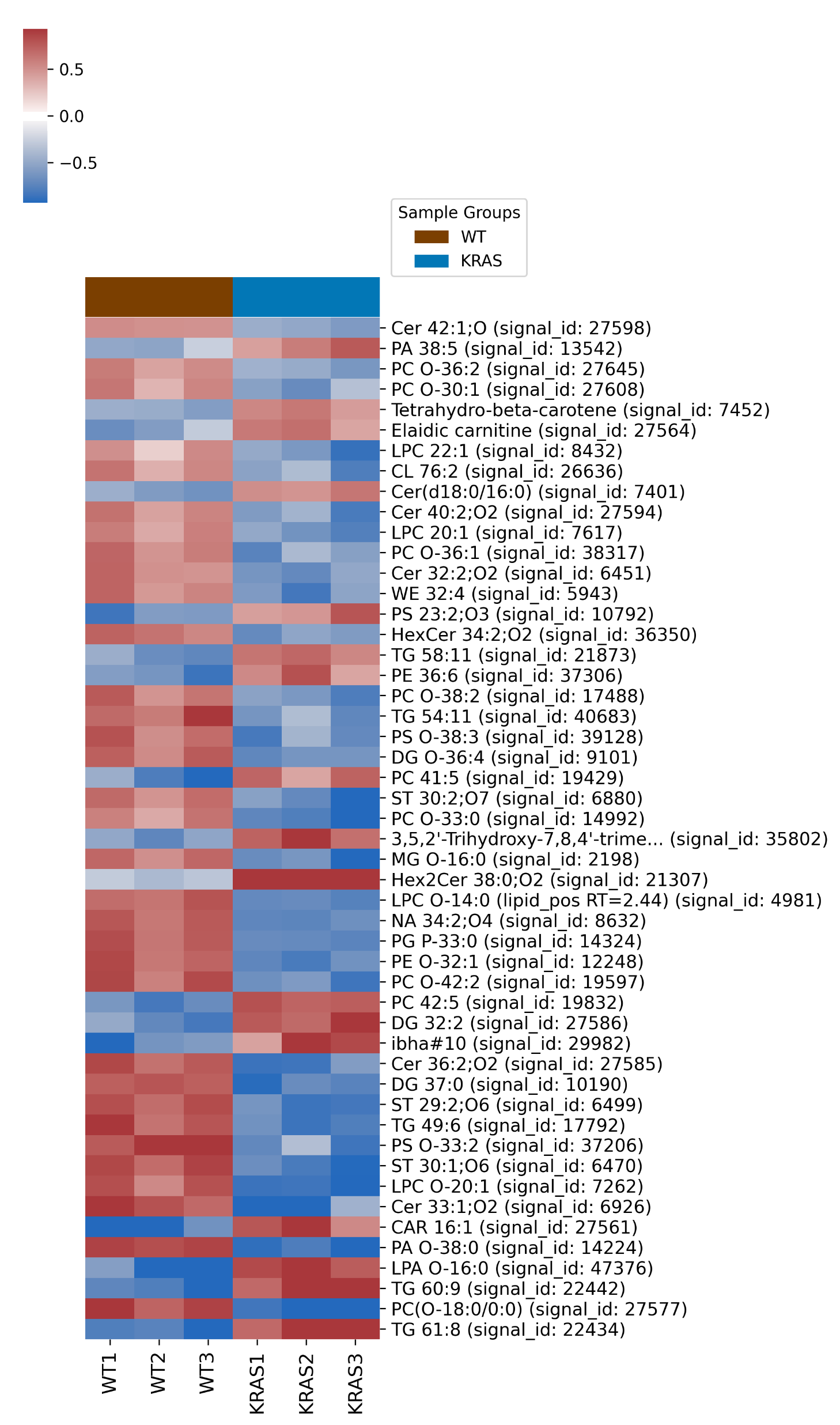
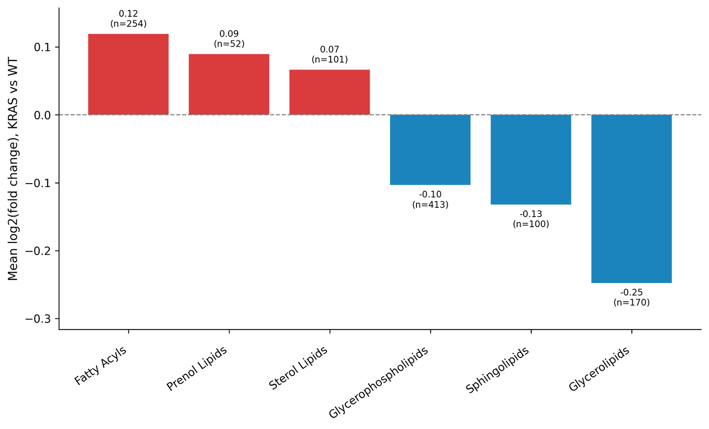
KRAS Knockdown Induces Broad Membrane Lipid Remodeling
Membrane lipid classes show coordinated shifts following KRAS knockdown, including reductions in sphingolipids and glycerophospholipids alongside increases in sterol- and fatty acyl-associated lipids, consistent with altered membrane organization and microdomain composition.
Heatmap clustering (left) shows again a clear separation between KRAS and wild-type samples, while fold-change analysis highlights coordinated increases and decreases across the surface proteome in the boxplot (right).
Coordinated Protein–Lipid Remodeling at the Cell Surface
Membrane proteins do not function independently of their surrounding lipid environment. Structural lipids such as sphingolipids, sterols and glycerophospholipids organize the membrane into specialized microdomains that regulate receptor localization, signaling activity, membrane fluidity and protein stability.
Integrated analysis of matched Cell Surface Proteomics and Cell Membrane Lipidomics data revealed biologically coordinated changes between altered surface proteins and membrane lipid class composition.
Specific examples of protein–lipid associations included:
- FLOT1 associated with sphingolipid- and sterol-rich membrane rafts
- FOLR1 associated with GPI-anchored raft localization
- SLC7A11 linked to glycerophospholipid redox balance
- TROP2 associated with phospholipid-mediated signaling pathways
Together, these findings suggest that KRAS knockdown drives coordinated remodeling of both membrane protein composition and the lipid microenvironments that regulate receptor localization and signaling behavior.
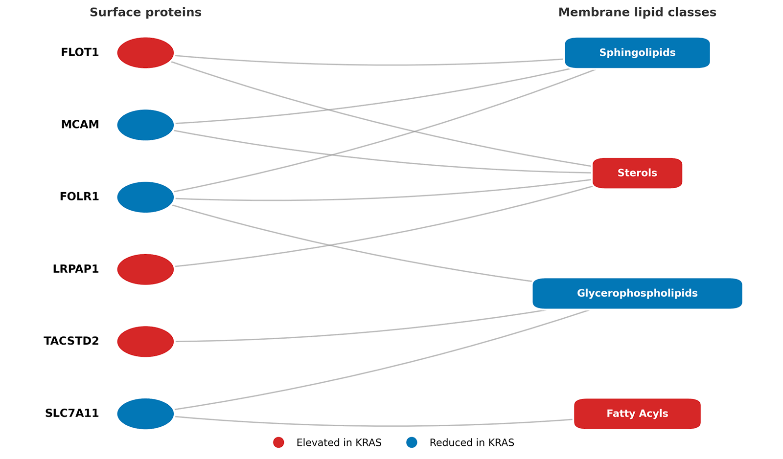
Coordinated Remodeling of Cell Surface Proteins and Membrane Lipid Classes in KRAS-Knockdown Cells
The integrated dataset supports KRAS-dependent rewiring of surface transport and signaling, including reduced nutrient transport and adhesion, remodeling of lipid raft-associated signaling domains, emergence of alternative surface proteins, and coordinated protein–lipid membrane restructuring. Cell surface proteins (left) are linked to membrane lipid classes (right) based on reported biological associations. Colors indicate direction of change in KRAS-knockdown HCT-116 cells (red = increased, blue = decreased), and links represent known protein–lipid relationships.
Applications of Cell Surface Multi-Omics
Panome Bio’s Cell Surface Proteomics and Cell Membrane Lipidomic can support among others:
- Cell surface target discovery
- Antibody Drug Conjugate and CAR-T target characterization
- Membrane signaling studies
- Lipid raft and receptor organization analysis
- Cancer membrane biology research
- Drug response profiling
Cell Surface Omics
Dive into our Data Report for a deeper biological interpretation of membrane remodeling.
Coordinated protein and lipid remodeling in KRAS-KD HCT-116
Explore the Application Note to for a better understanding of the full dataset and protein–lipid associations.
Surface Proteomics for Smarter Drug Target Discovery
Read the blog for a broader overview of how surface omics reveals signaling rewiring in cancer
Explore Cell Surface Multi-Omics with Panome Bio
Panome Bio’s Cell Surface Multi-Omics platform combines matched membrane proteomics and lipidomics to characterize coordinated remodeling at the cell surface. The platform enables simultaneous investigation of surface receptor expression, membrane lipid composition, protein–lipid coupling and membrane microdomain organization across cancer, immunology and cell signaling applications.
Contact us to start your project.